Bulletin E0922
Functional Anatomy of the Horse Foot
DOWNLOAD
February 12, 2016 - Melvin Bradley
 Print
Print Email
EmailThe hoof is composed of the wall, sole and frog. The wall is simply that part of the hoof which is visible when the horse is standing. It covers the front and sides of the third phalanx (coffin bone). The wall is made up of the toe or the front part; the quarters or sides and the heels.
Whenever the foot is lifted off the ground, the sole, frog, bars of the wall and collateral grooves are visible (Figure 1).
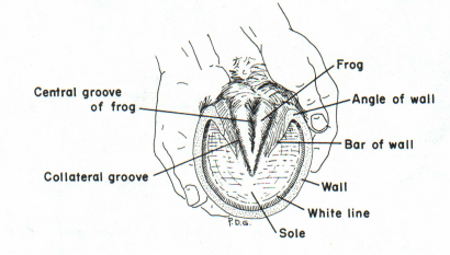
Figure 1.
The wall of the hoof is composed of a horny material which is produced continuously and must be worn off or trimmed off. The hoof wall does not contain blood vessels or nerves. In the front feet, the wall is thickest at the toe; in the hind feet there is less difference in the thickness of the hoof wall.
The frog is a wedge-shaped mass which is quite elastic. Its role will be discussed later.
Lateral cartilages extend back and up from the inner and outer sides of the third phalanx. These cartilages are flexible but as the horse ages they are usually replaced by bone (Figure 2).
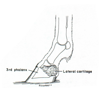
Figure 2.
The digital cushion is a mass of flexible material that contributes to the formation of the heels. This structure is very important as it is one of the primary shock absorbers of the foot.
A small bone, the navicular bone, is located between the second and third phalanges and above the deep flexor muscle tendon (Figure 3).
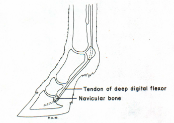
Figure 3.
As weight is placed on the hoof, it is transmitted through the phalanges to the wall and onto the digital cushion and frog. Normally the frog makes contact with the ground first. As the frog presses up on the cushion, the digital cushion is flattened and is forced out against the lateral cartilages. The frog also is flattened and tends to push the bars of the wall apart (Figure 4).
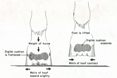
Figure 4.
When the foot is lifted, the above structures return to their original position. By placing the foot on the ground, blood is forced from the foot due to the pressure and change in shape of the digital cushion and frog. The pressure and change in shape compress the veins in the foot. More blood then fills the veins when there is no more compression that is when the foot is lifted. Therefore, the movement of these structures in the hoof acts as a pump. Exercise increases the blood circulation in the foot and favors good hoof growth. Lack of exercise, dryness of the horny wall and poor nutrition inhibits the rate of hoof growth. It is very important to realize that the sole normally does not contact the ground. The wall, bars and frog are the weight bearing structures of the foot (Figure 1).
The hoof wall grows at about the rate of three-eighths inch per month. The coronet is a ring which encircles the foot at the junction of the skin and hoof wall. Immediately distal to the coronet, new layers of hoof wall are produced.
The hoof wall is covered with material which prevents evaporation of moisture. When the material is deficient, the hoof wall becomes dry and excessive flaking and cracking may occur. A good hoof paint aids in preventing excessive drying.